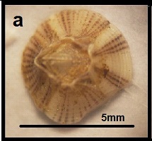
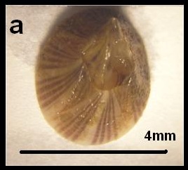
Purple Acorn Barnacle (Balanus amphitrite)
Table of Contents
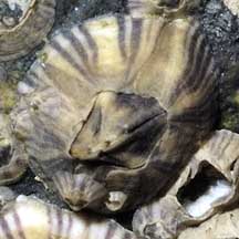
What are barnacles?‘‘A animal fixed by its head and kicking the food into its mouth with its legs” T.H. Huxley (English biologist, 1825 -1925)
General name
English: barnacle
Chinese: 藤壶
Malay: teritip
Arabic: البَرْنَقيل
Japanese: ふじつぼ
General characteristics
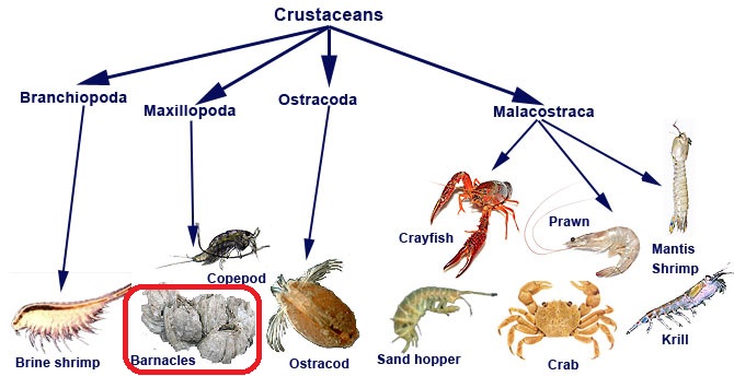 |
| Barnacles are relatives of the better known crustaceans i.e. crabs and shrimps. Pictures edited and taken with permission from Marine Education Society of Australasia (MESA) |
-they are crustaceans
- belong to the same group of animals (Subphylum Crustacea) including crabs, lobsters, crayfish, shrimp, krill (Solomon et al, 2008). All of them possess:
- two-parted (biramous) appendages
- segmented body parts: head, thorax and abdomen
- larval (naplius) stage in their life cycle
- calcareous shell: wall plates for barnacles, carapace for crabs and other crustaceans
- shell for protection, prevent dessication and communication (some crustaceans)
- belong to the same group of animals (Subphylum Crustacea) including crabs, lobsters, crayfish, shrimp, krill (Solomon et al, 2008). All of them possess:
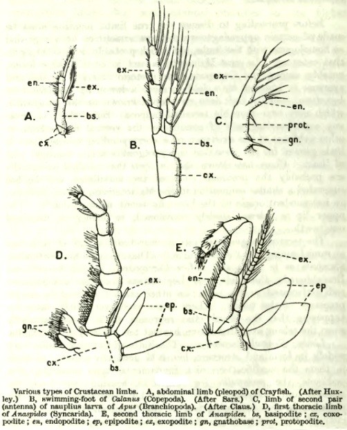 |
| Homology of biramous appendages of crustaceans. FIgure taken with permission from E. Ray Lankester |
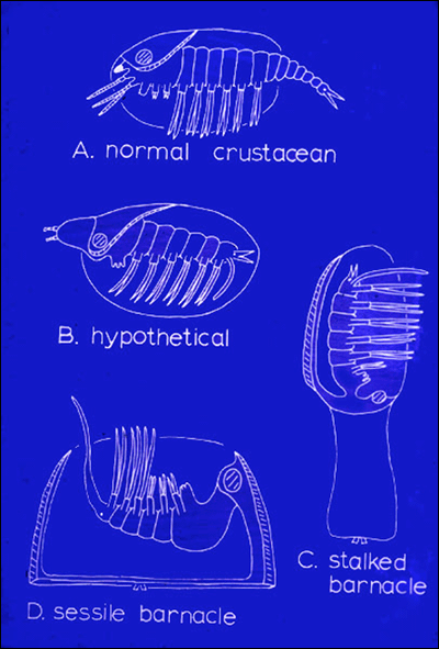 |
| Cartoon drawing showing homology of crustacean appendages. Figure adapted with permission from P.S. Rainbow (1984) |
-termed cirripeds
- found in Infraclass Cirripedia. Cirri in Latin, means "curled'', -pediameans legs (Solomon et al, 2008).
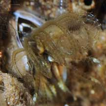 |
| Curled cirri of a typical barnacle. Picture taken with permission from Ria Tan |
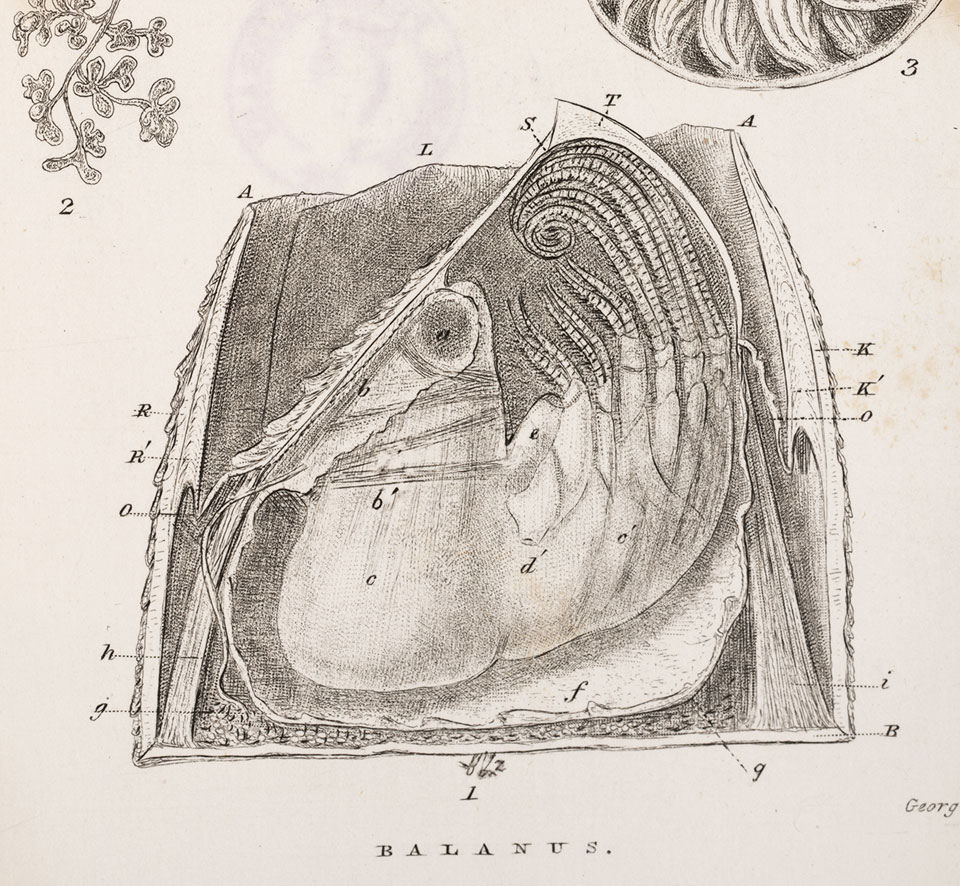 |
| Charles Darwin's illustration of the morphology of an acorn barnacle; with an enlarged longitudinal section through the shell and sack, with the right-hand scutum and tergum and right-hand half of shell and basis removed, exhibiting the body of the animal not in section. The cirri are exhibited only on one side. A, A, orifice of shell, within which lies the operculum formed by a pair of scuta (S), and pair of terga (T). B, basis (homologically the anterior end of the shell). K, carina of shell (or dorsal valve or compartment of shell). K´,sheath of carina. L, lateral compartment of shell. The carino-lateral compartment is hidden by the scutum and tergum. R, rostrum of shell (or ventral valve or compartment of shell). R´, sheath of rostrum. O, O, opercular membrane, connecting the opercular valves with the overhanging basal edge of the sheath. S, scutum. Picture taken with permission from Darwin-Online |
Types of Barnacles
-have different adult forms
- acorn or rock barnacles (Order Sessila)
- gooseneck or stalked barnacles (Order Pedunculata)
- parasitic barnacles (Superorder Rhizocephala)
 |
| Various forms of barnacles: acorn barnacles (left), goose barnacles (center) and parasitic barnacle(right). Pictures taken with permission from Wild Singapore and Wikipedia Commons |
Reproduction and Life History
-mostly hermaphrodites
- possess both male and female reproductive organs (Charnov, 1987).
- allow self-fertilization to produce offspring when there are no other barnacles nearby (Barnes & Crisp, 1956)
- if two barnacles happen to be close to one another, one will protrude a long penis into the other barnacle (pictured as below)
- penises can extend up to eight times its own body length (Darwin, 1854)
- have the highest penis to body length ratio amongst all animals (Neufeld & Palmer, 2009)
- different gender individuals are thought to be evolved from hermaphrodite ancestors (Charnov, 1987; University of Pittsburg, 2011)
- different life stages in a life cycle
- metamorphic life cycle (Zvyagintsev & Korn, 2003)
- starts out as a free-swimming larva, called a nauplius, undergoes six stages of naupliar growth;
- grows a pair of shells around its body now called a cypris larva;
- head of cypris larva attaches itself permanently to a hard structure;
- cypris now sessile, starts to develop into an adult: It casts off its shells and begins to secrete several calcareous plates;
- adult barnacle now releases barnacle glue
- metamorphic life cycle (Zvyagintsev & Korn, 2003)
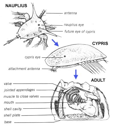 |
| Different life stages of an acorn barnacle and its anatomical description. Picture adapted from Lippson & Lippson (1984), awaiting permission |
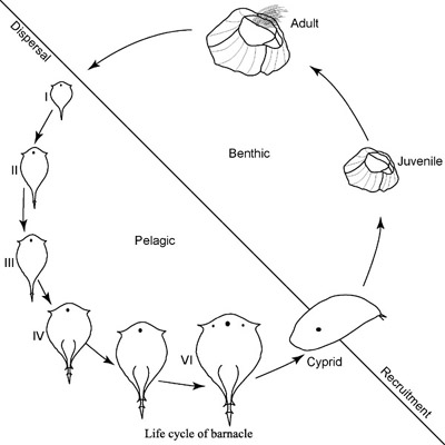 |
| Stages in a life cycle of a typical Balanomorph barnacle (Family Balanidae). Figure (awaiting permission) from National Institute of Oceanography |
-adult attaches to hard substratum with cement-like secretion
- similiar to the proteins of humans blood clot (platelets) (Dickinson et al, 2009)
- protein fibres released from cement ductsjunctions) between base-plate and wall/lateral plates
- Factor XII that induces release of platelet also found in barnacles
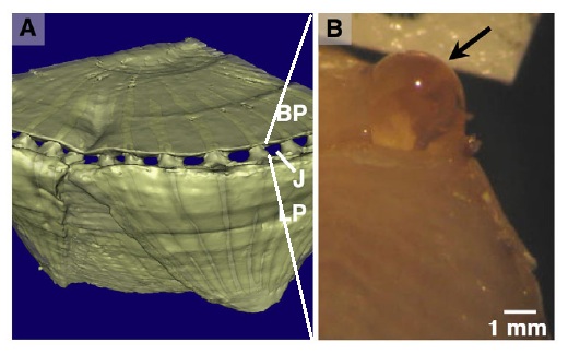 |
| Figure (A) X-ray microtomograph of a live barnacle (Amphibalanus amphitrite) showing the junction (J) of the base plate (BP) and lateral plates (LP), where cement is released through ducts during growth and Figure (B) showing a cement duct releasing cement for attachment. Figures taken with permision from Dickinson et al (2009) |
Feeding Habits
- feed when submerged or exposed to water (during high tides)
- operculum opens and cirri exposed capture food
- cirri retract into shell and operculum is closed when not exposed to water (exposed to air during low tide)
- capture prey by grabbing or filtering small particles
- uses feet-like appendages (cirri) which are attached to their limbs
- cirri placed perpendicular to the direction of water currents
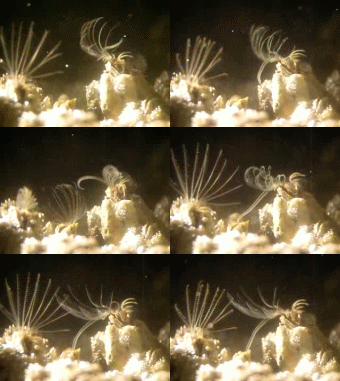
- grabbing: cirri swiped in and out of oral cavity at high velocity, 'grabbing' food particles in a repetitive manner (Crisp & Southward, 1961).
 |
| Video showing cirri of purple acorn barnacles (Balanus amphitrite) feeding on non-visible particles in water |
- filtering: when water current is slow, cirri are placed above the oral cavity (at different directions) similar to a mesh trap, retraction of cirri into oral cavity is slow (Crisp & Southward, 1961).
 |
| FIlter feeding of an acorn barnacle |
- General Food Chain
- feed on small and micro-sized particles e.g. bacteria, plankton, waste nutrients, etc.
- being predated by gastropods (snails and slugs) and other crustaceans (e.g. crabs)
- dog whelks (Nucella lapillus) are carnivorous sea snails
- use special drilling mouth parts (radula) to bore through the shells of barnacles (Largen, 1967)
- digestive enzymes are injected through the hole and the resulting liquid food sucked
- crabs: use larger pincers to crab the wall plates of barnacles and feed on them (Watch video below)
- dog whelks (Nucella lapillus) are carnivorous sea snails
 |
| Illustration showing a typical food chain involving barnacles. Illustration taken with (awaiting permission) from Young Peoples Trust for the Environment, UK |
Video showing a red crab (Gecarcoidea natalis) feeding on barnacles underwater. Video courtesy of Youtube user (tigertensing)
Habitat Range & Distribution
- only in marine environments: intertidal regions (beaches, mangroves, rocky shores), some deep water species
- attachment on any hard substratum including rocks, tree trunks, floating debris, buoys, ship hulls, etc. Some specialist species attach on turtles and whales
- worldwide distribution
- In Singapore:
- all coastal areas on mainland and offshore islands
- various species (Jones et al, 2000)
Biofouling
- biofouling: undesirable and gregarious attachment on objects
- barnacles fouling found on
- many human structures (buoys, ship hulls, harbors stilts, etc.)
- debris (both floating and non-floating)
- floating debris poses threat to introduction of exotic species(by ocean circulation to remote islands and shores around the world (Hansen, 1990)
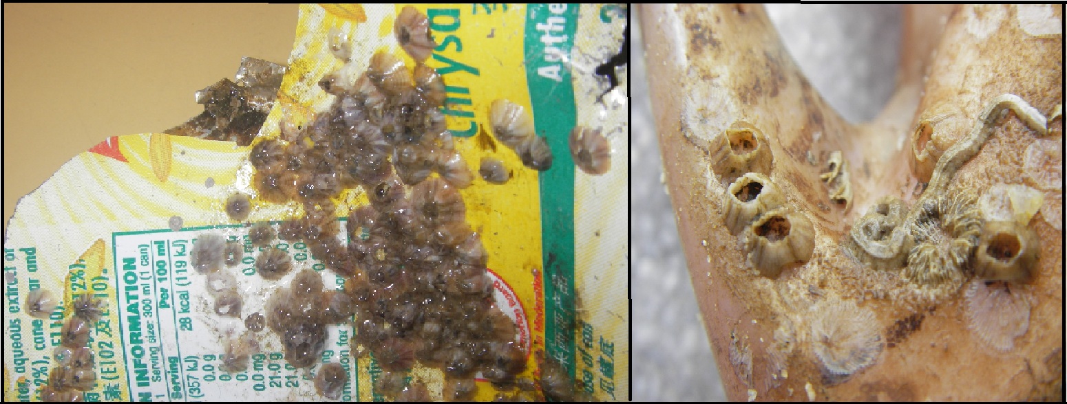 |
| Biofouling of barnacles on a aluminium tin can (left) and on a plastic debris(right). Pictures taken with permission from Leong Chin Rick |
Charles Darwin's study on the Purple Acorn Barnacle
(1) Description of Balanus amphitrite
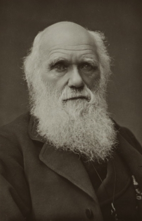 |
| Charles Darwin (1809-1882) was the Father of Barnacle Taxonomy. Picture taken with permission from Darwin-online |
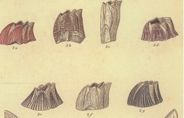 |
| Figure 2a: Darwin's illustration of Balanus amphitrite and 2b to 2g showing few varieties of the species. Figure adapted with permission from Darwin-Online |
- Charles Darwin (1854) studied barnacles and their taxonomy for eight years from 1846 to 1854.
- Initially described Balanus amphitrite as multivariate species
- named B. amphitrite and identified other nine varieties namely Balanus amphitrite var. communis, venustus, pallidus, niveus, modestus, stutsburi, obscurus, variegatus, and cirratus.
- All of them shared the following morphological and anatomical characteristics of the following:
Scutum internally with a prominent broad adductor ridge"
| Balanus amphitrite. a. top view and b. internal view of tergum (top-right) and scutum (bottom-left) and c. external view of scutum and d. internal view of scutum.Pictures taken with permission from Leong Chin Rick |
- After Darwin, other scientists found some of the varieties to be of the same species
- supports Darwin's theory of evolution by natural selection ; 'no two individuals in a species are morphologically alike'
(2) Description of Distribution
- Darwin (1854) found B. amphitrite to be commonly found in warmer temperate areas: tropical seas and intertidal areas.
- extremely common; Mediterranean, Smyrna(now Izmir, Turkey), Sicily (Italy), Coast of Portugal, West Coast of Africa, River Gambia, West Indies, Demerara (now Guyana), (Kwa-Zulu) Natal (South Africa), Madagascar, Red Sea, Mouth of the Indus(Indus River mouth), Ceylon(now Sri Lanka), Philippine Archipelago, East Indian Archipelago, Pacific Ocean, east coast of Australia, New Zealand; extremely common on ships' bottoms; often attached to floating timber, canes
- Often associated with B. tintinnabulum; attached to pebbles and various shells
Refer to 'Species Distribution' and 'Invasion' segments below for further information of this species' distribution
For further reading of Charles Darwin's study on barnacles, please read his monograph here. Refer to page 240 of the monograph for Charles Darwin's description of Balanus amphitrite.
Origin of Name (Etymology)
- Scientific (Binomial) Name: Balanus amphitrite Darwin 1854 (= Amphibalanus amphitrite Pitombo 2004)
- Balanus = chestnut or acorn in Latin (Da Costa,1778)
- amphitrite= wife of King Poseidon in Greek Mythology; goddess of the oceans
- Revision of genus name from Balanus to Amphibalanus, now known as Amphibalanus amphitrite (Pitombo, 2004)
- Amphibalanus is combinition of terms amphitrite and balanus
- revision due to introduction of new monophyletic g Amphibalanidae from original Balanidae
- Old Scientific (Binomial) Synyonyms: Lepas radiata (Woods, 1815)
- Vernacular Names: striped barnacle, the purple acorn barnacle and Amphitrite's rock barnacle
- ‘striped barnacle’ because its wall plates have longitidunal coloured striation
- Current Status of Scientific Name
- usage of Barnacle amphitrite still exist (Clare & Hoeg, 2008; Carlton & Newman, 2010).
- Clare and Hoeg (2008) supported retention of B. amphitrite due to “scarce” referencing of A. amphitrite, and outright critism of Pitombo (2004)’s methology of phylogenetic revision.
- Carlton & Newman (2010), refuted such claims stating that names changes, especially to non-systematists is often slow and does not reflect rejection from the scientific community and supports Pitombo’s revision.
- usage of Barnacle amphitrite still exist (Clare & Hoeg, 2008; Carlton & Newman, 2010).
Diagnosis
Balanus amphitrite tends to be confused with Balanus reticulatus
- Simple Identification (for non-taxonomists)
- Not accurate due to morphological variation and habitat variation
- Simple terminologies adapted from Chan et al (2009)
| Balanus reticulatus (= Amphibalanus reticulatus) |
Balanus reticulatus (= Amphibalanus reticulatus) |
||||
|
|
||||
| Brown and thin-line striations |
Purple and thick-line striations |
||||
| Transverse striations across wall plates |
No transverse striations across wall plates |
||||
| Rough wall plates |
Smooth wall plates |
- Specific Identification (for taxonomists)
- accurate but requires microscopy skills to identification
- Kindly refer to Fox's Guide to Invertebrate Anatomy Online for reference of morphological terms of a Balanomorph barnacle.)
Shell: Wall of six plates, smooth; parietes with a single row of tubes with or without transverse septa; radii solid, transverse teeth on sutural edge with denticles on lower side only (Pitombo, 2004;Chan et al, 2009).
Basal surface:Basis tubiferous, tubes in a single layer(Pitombo, 2004;Chan et al, 2009).
Operculum: Scutum with a conspicuous adductor ridge. Tergum with spur having abrupt change in the direction of growth lines, with spur and furrow margins coincident, basal margin with well-developed depressor muscle crests projecting beyond border (Pitombo, 2004;Chan et al, 2009).
Oral appendages: Second maxilla with anterior margin of distal lobe having smooth, acuminate setae with enlarged and modified tips.((Pitombo, 2004;Chan et al, 2009).
Cirri (feet-like appendages): Cirrus III with inner face of endopod with pinnate setae rarely with bifurcate (complex) setae. CirriIV-VI with erect hooks below posterior angles of distal articles of rami (Pitombo, 2004;Chan et al, 2009).
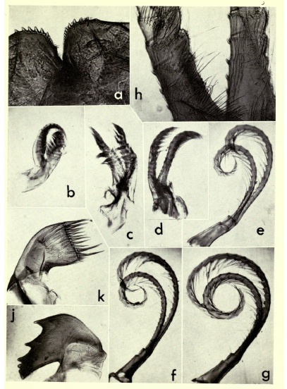 |
| Cirri I, II, III, IV, V and VI (Figures b- g) of Balanus amphitrite, Figure taken with permission from British Natural History Museum Library |
Species Distribution
- widely distributed (cosmopolitan) tropical and subtropical species (Newman & Ross, 1976)
- in subtropical areas, B. occurs abundantly in the intertidal zone of sheltered coasts, usually below the mean seawater level
- occurs intertidally, often in brackish waters (Henry & Mclaughin, 1975)
- able to withstand low salinity levels (Southward,1975; Utinomi, 1960)
- often abundant in habitats exposed to physical stress and pollution, including oil covered areas (Lipkin & Safriel, 1971)
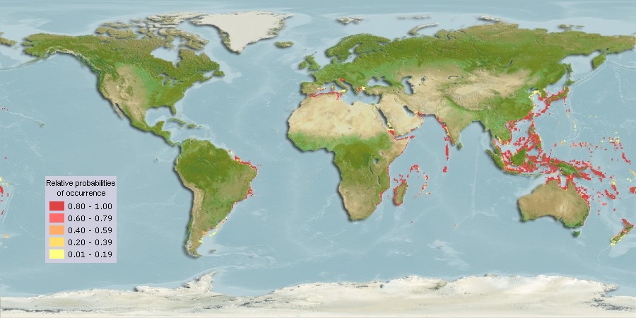 |
| Computer generated map for Balanus amphitrite as of 2010. Map taken with permission from Aqua Maps |
- Balanus amphitrite was found to be originated from the Southwestern Pacific and Indian Oceans from discovery its fossils (Zullo 1963).
- found in Singapore as well (Jones et al, 2000).
Ecology
Invasion
- expected to be confined to warm and tropical waters but found to be invasive in several colder and temperate countries including
- Northern America (United States of America),
- Southern America (Argentina)(Oresanz et al., 2002)
- Europe (Germany, Belgium) (Wiegemann, 2008; Kerckhof, 2002)
- Northeast Asia (Japan and Hong Kong) (Qiu, 1999)
- Oceania (Australia ands New Zealand) (Foster, 1978)
- invasion may come about from biofouling on ship hulls and floating debris travelling around oceans (Lulito, 2007)
- Current IUCN (International Union for Conservation of Nature) Threat Status: Unknown
Interspecies Competition
- Interspecies competition: individuals of different species compete for the same resource(e.g. food, living space) in an ecosystem.
- Between Balanus amphitrite and polychaete worm (marine worm Pomatoleios kraussii)
- heavy mortality of B. amphitrite is associated with high density of P. kraussii in same area of attachment (Mohammad, 1975)
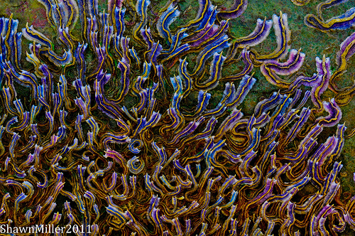 |
| Balanus amphitrite's competitior for space and resources. Photo taken with permission from Shawn Miller. |
Taxonavigation
(Definition of 'taxonavigation': click here)Kingdom Animalia -- Animal, animals, animaux
Phylum Arthropoda -- arthropodes, arthropods, Artrópode
Subphylum Crustacea Brünnich, 1772 – crustace, crustáceo, crustacés
Class Maxillopoda Dahl, 1956
Subclass Thecostraca Gruvel, 1905
Infraclass Cirripedia Burmeister, 1834 -- barnacles, bernacles
Superorder Thoracica Darwin, 1854 -- barnacles
OrderSessilia Lamarck, 1818 -- sessile barnacles
Suborder Balanomorpha Pilsbry, 1916 -- acorn barnacles
Superfamily Balanoidea Leach, 1817
Family Balanidae Leach, 1817/ Amphibalanidae Pitombo, 2004)
Genus Balanus Da Costa, 1778 /Amphibalanus Pitombo, 2004)
Species Amphitrite Darwin, 1854
Classification adapted from ITIS and Pitombo (2004).
Type Information
- Type (in Biology): a specific specimen or group of specimens of an organism to which the scientific name of that organism is formally identified as.
- The reason for such designation is to avoid a confusion about applying names if someone revises the genus or species.
- Darwin did not designate type specimen (and a holotype) for B. amphitrite and its varieties (Yamaguchi, 1980)
- Harding (1962) assigned lectotypes (lectotype: a specimen chosen as the type of a species or subspecies if the original author of the name fails to designate a type) for Darwin’s dried specimens
- lectotype was originally found on a piece of bamboo (Registration No. :B. M. 40.9.15.17, locality Natal (South Africa), Dr. Krauss)
- lectotype was dissected, now kept separately as:
- remaining shell on the bamboo registered as B.M. 1961. 12. 6. 1c
- mounted operculum valve as B.M. 1961.12.6. 1a
- animal parts as B.M. 1961.12.6. 1b (Harding, 1962).
Link to Other Species Pages
Marine Species
Exotics Guide
Sea Life Base
Encylopedia of Life
Cirripedia My Species
Introduced Marine Species of Hawaii
Literature and References
Anderson, D. T. 1980. Barnacles - structure, function, development and evolution. Chapman and Hall London. 357 pp.
Barnes, H. & D.J. Crisp, 1956. Evidence of self-fertilization in certain species of barnacles. J. Mar. Biol, 144: 235-239.
Chan, B.K.K., Prabowo, R.E and Lee, K.S. (2009), Crustacean fauna of Taiwan: Barnacles, Volume I- Cirripedia: Thoracica excluding the Pyrgomatidae and Acastinae. National Taiwan Ocean University, Keelung. 297pp.
Charnov, E.L. 1987. Sexuality and hermaphroditism in barnacles: a natural selection approach. In Southward A.J. (ed.) Crustacean issues 5, Barnacle biology. Rotterdam: A.A. Balkema, pp. 89–104.
Clare, A.S. & Høeg. J.T. 2008. “Balanus amphitrite or Amphibalanus amphitrite? A note on barnacle nomenclature.” Biofouling 24(1): 55-7.
Computer Generated Map for Balanus amphitrite (un-reviewed). www.aquamaps.org, version of Aug. 2010. Web. Accessed 14 Nov. 2011.
Crisp, D. J. & A. J. Southward, 1961. Different types of cirral activity of barnacles. Phil. Trans. Roy. Soc., Scr. B, 243 : 271-307.
Darwin, C. R. 1854. Living Cirripedia, The Balanidæ, (or sessile cirripedes); the Verrucidæ. London: The Ray Society. Volume 2 Text Image Text & image F339.2
Dickinson, G. H.,Vega, I.E., Wahl, K. J., Orihuela, B., Beyley, V., Rodriguez, E.N., Everett, R. K., Bonaventura, J. & D. Rittschof, 2009. Barnacle cement: a polymerization model based on evolutionary concepts, Journal of Experimental Biology, 212(21): 3499-3510.
Foster, B.A. 1978. The marine fauna of New Zealand: Barnacles (Cirripedia, Thotacica). Memoirs of the New Zealand Oceanographic Institute 69: 1-160.
Hansen, J. 1990. Draft position statement on plastic flotsam in marine environments. Fisheries. 15: 16–17.
Harding J. P. 1962. Darwin's type specimens of varieties of Balanus amphitrite. Bull. Br. (Natn Hist.) Zool., 9(7): 273-296
Henry, D. P. & Mclaughlin, P. A. 1975. The barnacles of the. Balanus amphitrite complex (Cirripedia, Thoracica). Zoologische. Verhandelingen, 141, 1–254
Jones, D.S, Hewitt, M. A. & A. Sampley. 2000. A Checklist Of The Cirrepedia of South China Sea. Raffles Buletin of Zoology. 8: 203-307.
Kerckhof, F. (2002). Barnacles (Cirripedia, Balanomorpha) in Belgian waters, an overview of the species and recent evolutions, with emphasis on exotic species. Bull. Kon. Belg. Inst. Natuurwet. Biologie, 72(Suppl.): 93-104
LaBarbera, M. 1984. "Feeding currents and particle capture mechanisms in suspension feeding animals". American Zoologist, 24 (1): 71–84.
Largen, M. J. 1967. The diet of the dog-whelk, Nucella lapillus (Gastropoda Prosobranchia). Journal of Zoology, 151(1): 123-127
Lulito, C. 2007. Distribution, abundance and reproduction of the Indo-Pacifric acorn barnacle Balanus amphitrite (Crustacea: Cirripedia). J. Mar. Biol. Ass. U.K., 87: 723-727.
Lipkin, Y. & Safriel, U. 1971. Intertidal zonation on rocky shores at Mikhmoret (Mediterranean, Israel). The Journal of Ecology, 59:1-30.
Lippson, A.J. & Lippson, R.L. 1984. Life in the Chesapeake Bay. Johns Hopkins University Press. 304pp.
Mohammad, M.B.M. 1975. Competitive Relationship Between Balanus amphitrite and Pomatoleios kraussiiwith Special Reference to their Larval Settlement.
Hydrobiologia, 46:1-15.
Neufeld, C. J. & A. R. Palmer. 2008. Precisely proportioned: intertidal barnacles alter penis form to suit coastal wave action. Proceedings of the Royal Society B-Biological Sciences, 275:1081-1087.
Newman, W. A. & Ross, A. 1976. Revision of the Balanomorph barnacles including a catalogue of the species. San Diego Society of Natural History Memoirs, 9: 1–108.
Orensanz, J.M., Schwindt, E., Pastorino, G., Bortulas, A., Casas, G., Darrigan, G., Elías, R., Gappa, J.J.L., Obenat, S., Pascual, M., Penchaszadeh, P., Piriz, M.L., Scabarino, F., Spivak, E.D. and E.A. Vallarino. 2002. No longer the pristine confines of the world ocean: a survey of exotic marine species in the southwestern Atlantic. Biological Invasions, 4: 115-143.
Pitombo, F. B. 2004. Phylogenetic analysis of the Balanidae (Cirripedia, Balanomorpha). Zoologica Scripta, 33: 261–276.
Qiu, J.W. 1999. Tolerance of the barnacle Balanus Amphitrite amphitrite to salinity and temperature stress:effects of previous experience. Marine Ecology Progress Series. 188: 123-132.
Rainbow, P.S. 1984. An introduction to the biology of British littoral barnacles. Field Studies, 6: 1-51.
Solomon, E., Berg, l. & Martin, D. 2008. Biology. 8th ed. Brooks/Cole Publishing. 1234p.
Southward, A.J. 1975. Intertidal and shallow water Cirripedia of the Caribbean. Stud Fauna Curaqao Other Caribb Is1, 46: l-53.
University of Pittsburgh (2008, November 20). Two From One: Evolution Of Genders From Hermaphroditic Ancestors Mapped Out. ScienceDaily. Retrieved November 15, 2011, from http://www.sciencedaily.com/releases/2008/11/081120171328.htm
Utinomi, H.1960. On the world-wide dispersal of a Hawaiian barnacle, Balanus amphitrite hawaiiensis. Broch. Pac Sci,14: 43-50.
Wiegemann, M. 2008. Wild cyprids metamorphosing in vitro reveal the presence of Balanusamphitrite Darwin, 1854 in the German Bight basin, Aquatic Invasions, 3(2): 235-238.
Yamaguchi, T. 1980. A New Species Belonging to the Balanus amphitrite Darwin Group (Cirripedia, Balanomorpha).Journal of Paleontology, 54(5): 1084-1101.
Zvyagintsev, Y.A., & O.M. Korn. 2003. Life history of the barnacle Balanus amphitrite Darwin and its role in fouling communities of Peter the Great Bay, Sea of Japan. Biologiya Morya. 29: 50-58.
Zullo, V. A. 1963. A Preliminary Report On Systematics And Distribution Of Barnacles (Cirripedia) Of Cape Cod Region. Systematics-Ecology Program, Marine Biology Laboratory, Woods Hole, Massachusetts, 33 p.
.
HTML Comment Box is loading comments...
